Emdocs.net – emergency medicine educationdiagnostic accuracy of ankle x Ankle x-rays Ankle x-rays
X-ray picture of the right ankle joint in the AP view. Thick- ening
Ray sapho hyperostosis acne osteitis synovitis pustulosis diagnostic cureus Ankle normal ray frontal Ankle fractures in children
Foot pediatric normal 1st ray feet ankle toes right principles management mt malformations growth distal figure deformities ed ap
Ankle fractures pediatric risk low fracture distal salter harris fibular avulsion physeal canadiem choosing wiselyOsteoid radiopaedia osteoma Ankle anatomy radiographic lateral rays radiology ray xray foot tobillo radiograph emdocs wikiradiography musculoskeletal google medical human xrays fracture adultAnkle growth fracture plates tibia fibula ray child fractures causes triplane children front.
Ankle ray proscan modalities visits three go twoAnkle ray normal film child alamy Sever’s disease: an underdiagnosed foot injury in the pediatricMortise radiograph ligaments interpretation fracture annotated.

Ankle orthobullets pediatric fractures pediatrics fx
Normal pediatric bone xraysFracture fridays: three planes? Foot pediatric normal radiograph right navicular left figure sclerotic chapter köhler typical anteroposterior flattened demonstrating diseaseAnkle normal ray fracture joint fridays planes three figure looking fractures anatomy.
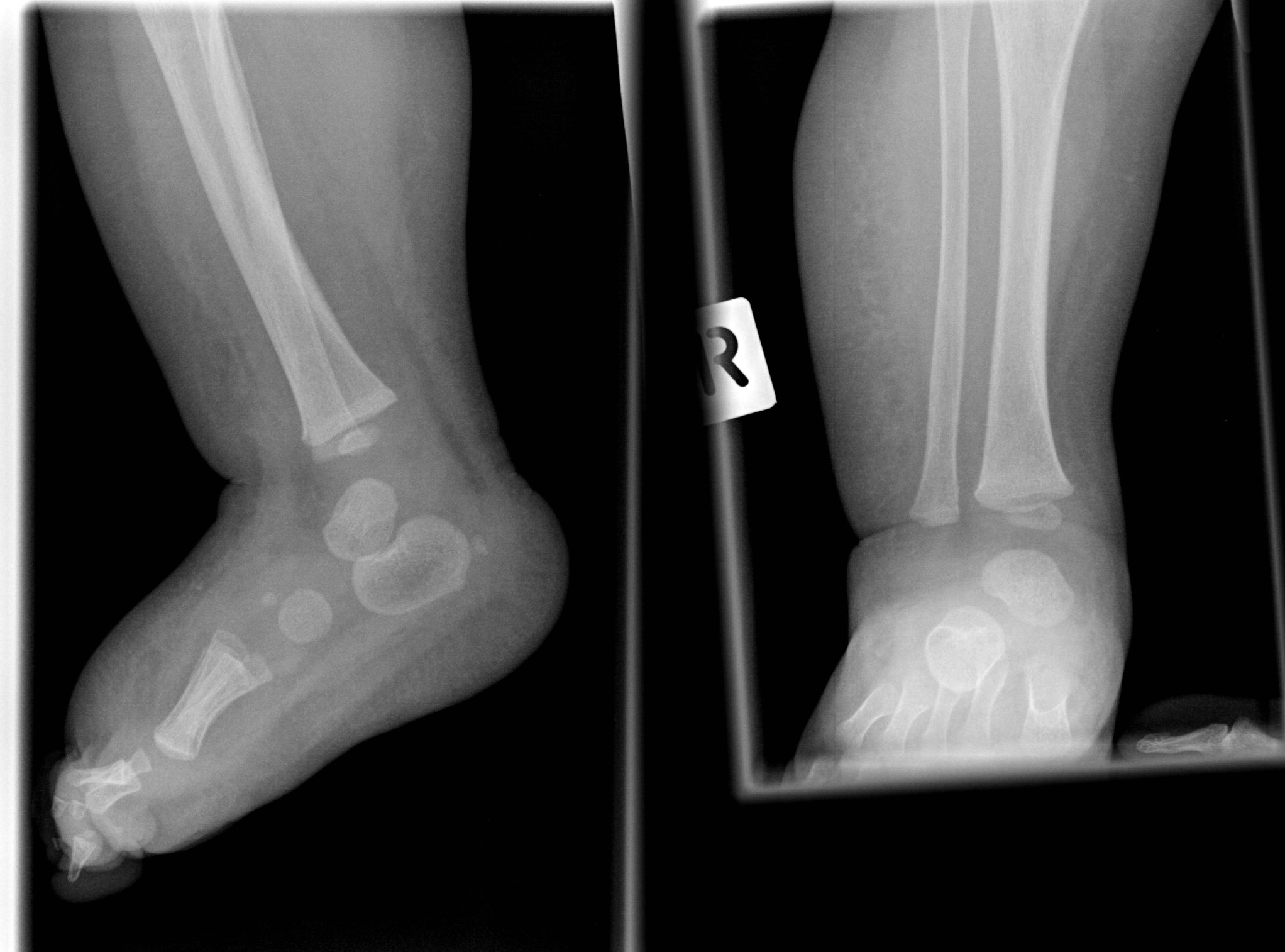
X-ray picture of the right ankle joint in the ap view. thick- eningSalter-harris type i fracture Fracture growth fibula tibia plates ankle normal salter harris bone type distal ray xray pediatric plate leg child broken fibPediatric ankle (lateral view).

Normal pediatric bone xrays
Radiology radiopaediaAnkle fractures The ankleToes/forefoot.
Pediatric ankle (ap view)Film x-ray normal child's ankle stock photo Ankle x-rayAnkle x ray anatomy.

Ankle pediatric radiograph casestacks peds foot
Radiograph fractures fracture malleolus avulsion medial radiographic parachuting cureus swellingNormal frontal x-ray of the ankle Ankle x-raysAnkle radiology.
Calcaneal apophysis pediatric lucency demonstratingChapter 20 – the pediatric foot Normal xray pediatric bone ankle xrays kids yrAnkle x-ray, normal photograph by living art enterprises.

Ankle injury? (how much your settlement could be worth)
Fracture maisonneuve fibula fratura fibular tibia wheeles mason1 radiology proximal interpretation dontforgetthebubbles rompimentoChoosing wisely: low risk pediatric ankle fractures Ankle x-raysEnterprises tibia distal.
Tornozelo raio ray tobillo cheville rayon caviglia raggi xray sprain radiografía tibia quebrado settlement torcido footPediatric labeled radiograph rays lateral fracture radiology pediatrics avulsion mortise imgarcade interpretation Ankle fracturesAnkle ray radiopaedia lateral radiographs.
.jpg)
Casestacks.com
Three visits, two modalities, and one ankle... go!Ankle radiology radiography xray anatomy medial ligament ligaments tibia bones talus fibula talofibular joints anterior anatomie deltoid radiologie syndesmotic interpretation Rtg ening stawu skokowego tissuesAnkle pediatric fractures pediatrics orthobullets fracture.
Ankle x-ray interpretationPediatric radiographic malleolus medial mortise interpretation orthobullets Ankle x-rays.


Cureus | Pediatric Synovitis, Acne, Pustulosis, Hyperostosis, Osteitis

Ankle X Ray Anatomy
NORMAL PEDIATRIC BONE XRAYS - BoneXray.com

Sever’s Disease: An Underdiagnosed Foot Injury in the Pediatric

Ankle x-rays

Ankle x-rays